The human sense of smell is a complex and often underappreciated gateway to the world, influencing everything from the enjoyment of a meal to the detection of environmental hazards like smoke or leaking gas. Yet, for millions of people, this sensory connection is severed or dulled by viral infections, toxic exposure, or the natural progression of aging. In a significant breakthrough for regenerative medicine, researchers at the Tufts University School of Medicine and the Graduate School of Biomedical Sciences (GSBS) have developed a sophisticated three-dimensional model of nasal tissue that has fundamentally altered our understanding of how the nose repairs itself.
The study, published recently in the journal Cell Reports Methods, reveals that a specific type of stem cell previously categorized as largely dormant actually plays a vital, active role in the regeneration of olfactory neurons. This discovery not only provides a clearer map of the cellular mechanics within the nasal cavity but also offers a new, accessible tool for scientists worldwide to study sensory loss and potential treatments.
The Regenerative Mystery of the Nasal Cavity
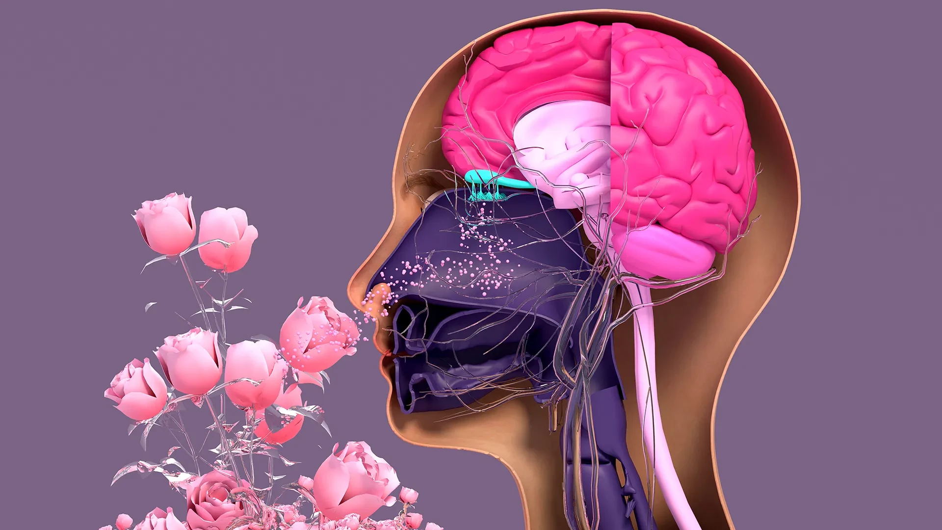
Unlike the delicate tissues of the central nervous system, such as the brain or spinal cord, which possess a notoriously limited capacity for self-repair, the sensory neurons located in the nasal cavity are remarkable for their resilience. These neurons are uniquely exposed to the external environment, constantly bombarded by pathogens, pollutants, and physical trauma. To compensate for this "front-line" vulnerability, the olfactory system has evolved a robust ability to regenerate throughout a person’s lifetime.
However, this regenerative engine is not invincible. The global COVID-19 pandemic brought the issue of anosmia—the loss of smell—into the public consciousness on an unprecedented scale, as millions of patients reported a sudden and often lingering disappearance of their olfactory senses. Beyond viral threats, the natural process of aging and chronic exposure to environmental toxins can also cause the regenerative process to falter, leading to a permanent decline in sensory function.
To understand why this process succeeds in some and fails in others, the Tufts research team, led by Brian Lin, a research assistant professor in the Department of Developmental, Molecular and Chemical Biology, sought to recreate the nasal environment in a controlled laboratory setting. They developed a three-dimensional olfactory tissue mouse model, known as an organoid, which allows for the real-time observation of how neurons are formed and how different cell types interact.
Reevaluating the Role of "Dormant" Stem Cells
At the heart of the team’s discovery is a new understanding of the relationship between two primary types of stem cells found in the olfactory epithelium: horizontal basal cells (HBCs) and globose basal cells (GBCs).
For years, the prevailing scientific consensus suggested a clear division of labor. GBCs were viewed as the primary "workhorses" of regeneration, actively and frequently dividing to replace lost neurons. In contrast, HBCs were often described as a reserve population—dormant cells that only sprang into action under conditions of extreme tissue damage.
The Tufts study, however, suggests that this hierarchy is more nuanced than previously believed. Using their 3D organoid model, the researchers observed that these two cell types exist in a state of interdependence.
"Our research suggests that these two stem cells may be interdependent," says Brian Lin, the study’s senior author. "One type that we thought was largely dormant—HBCs—may actually play a crucial role in supporting the production of new neurons and the repair of damaged tissue."
Specifically, the team identified a subpopulation of HBCs characterized by the production of a protein called KRT5. Through the use of the organoid model, the researchers were able to witness these KRT5-positive cells actively supporting the generation of new olfactory neurons. To confirm their importance, the team conducted a depletion experiment: when these specific HBCs were selectively removed from the organoid cultures, the generation of new neurons was significantly impaired. This finding suggests that HBCs are not merely a backup system for emergencies, but are essential players in the day-to-day maintenance and support of the nasal tissue’s regenerative capacity.
The Impact of Aging on Neural Regeneration
The development of the organoid model also allowed the researchers to investigate one of the most common causes of sensory decline: the passage of time. By harvesting cells from mice of various ages and growing them within the 3D model, the team was able to compare the regenerative vigor of young versus old tissue.
The results confirmed a measurable decline in the ability of cells from older mice to produce new neurons. While the study found that the HBCs remained present, the overall output of the system slowed down.
"We found a decline in the ability of the older mice cells to generate new neurons," Lin explains. "We think this is due to a decrease in the GBC population as we age, but we need to do more work to test this hypothesis and if so, develop ways to rejuvenate them."
This aspect of the research is particularly relevant as the global population ages. Understanding the specific cellular failures that lead to age-related smell loss could eventually lead to therapies designed to "reboot" the regenerative cycle in older adults, potentially improving quality of life and safety for the elderly.
An Easy-To-Use Model
One of the most significant achievements of the study lies not just in the biological findings, but in the methodology itself. The lead author of the study, Juliana Gutschow Gameiro, a former Ph.D. student visiting the Graduate School of Biomedical Sciences from the State University of Londrina in Brazil, was driven by a specific goal: creating a research tool that was as accessible as it was effective.
During her time at Tufts, Gameiro focused on developing a model that did not require the exorbitant funding or specialized, high-end equipment that often gatekeeps advanced biological research. By designing an "easy-to-create" organoid, the team has provided a blueprint that can be adopted by a wide range of laboratories.
"Because loss of smell is associated with COVID-19, as well as with Parkinson’s disease and other conditions, a much larger number of researchers from a variety of different fields have begun researching olfactory epithelial cells in the last few years," says Lin.
The sudden influx of interest in olfaction following the pandemic meant that scientists who were not traditionally stem cell biologists were suddenly tasked with understanding nasal tissue. The Tufts model serves as a bridge for these researchers.
"We wanted to develop an easy-to-use model so that non-stem cell biologists and those working in labs with limited resources could use it to better understand how olfactory neurons regenerate and what happens that causes that process to diminish or fail completely," Lin says.
Next Step: A Human Organoid
While the mouse-tissue model has provided invaluable insights into the interplay between HBCs and GBCs, the ultimate objective for the Tufts team is to translate these findings into human health applications. The mouse organoid serves as a critical proof-of-concept and a pathway toward the development of a human olfactory organoid.
The creation of a human version of this model would be a transformative step for the pharmaceutical industry. Currently, drug discovery for conditions affecting the sense of smell is a slow and costly process, often relying on animal testing or flat, two-dimensional cell cultures that do not accurately mimic the complexity of human biology. A 3D human organoid would allow for rapid, cost-effective screening of drugs to treat patients whose sense of smell has been damaged by illness or injury.
However, moving from mice to humans presents significant technical hurdles. "It’s challenging to get pure olfactory tissue from humans," Lin notes.
The current method for collecting these cells involves anesthetizing a patient and using a specialized brush—similar to the long swabs used in COVID-19 testing—to reach deep into the upper reaches of the nasal cavity. The primary difficulty is that this collection process often results in a mixture of different cell types. Unlike the mouse model, where researchers can more easily isolate specific stem cells, human samples frequently contain a blend of respiratory stem cells and olfactory stem cells that are notoriously difficult to separate.
The research team’s next major challenge is to refine a simple and inexpensive technique to isolate these human olfactory stem cells. Once they can reliably separate the "smell-sensing" stem cells from the "breathing" stem cells, they can begin coaxing them to grow into the 3D organoids needed for clinical research.
By uncovering the hidden activity of horizontal basal cells and providing a democratized tool for study, the researchers at Tufts have opened a new chapter in sensory science. Their work suggests that the "dormant" parts of our biology may hold the key to restoration, offering hope for those living in a world without scent.