In a significant advancement for sensory biology, researchers at Tufts University School of Medicine and the Graduate School of Biomedical Sciences (GSBS) have developed a sophisticated three-dimensional model to study the regeneration of nerve tissue within the human nose. The study, conducted alongside international colleagues, has led to a surprising discovery regarding the mechanisms of olfactory recovery: a specific type of stem cell, long categorized by the scientific community as largely dormant, appears to play a far more vital role in maintaining and restoring the sense of smell than previously understood.
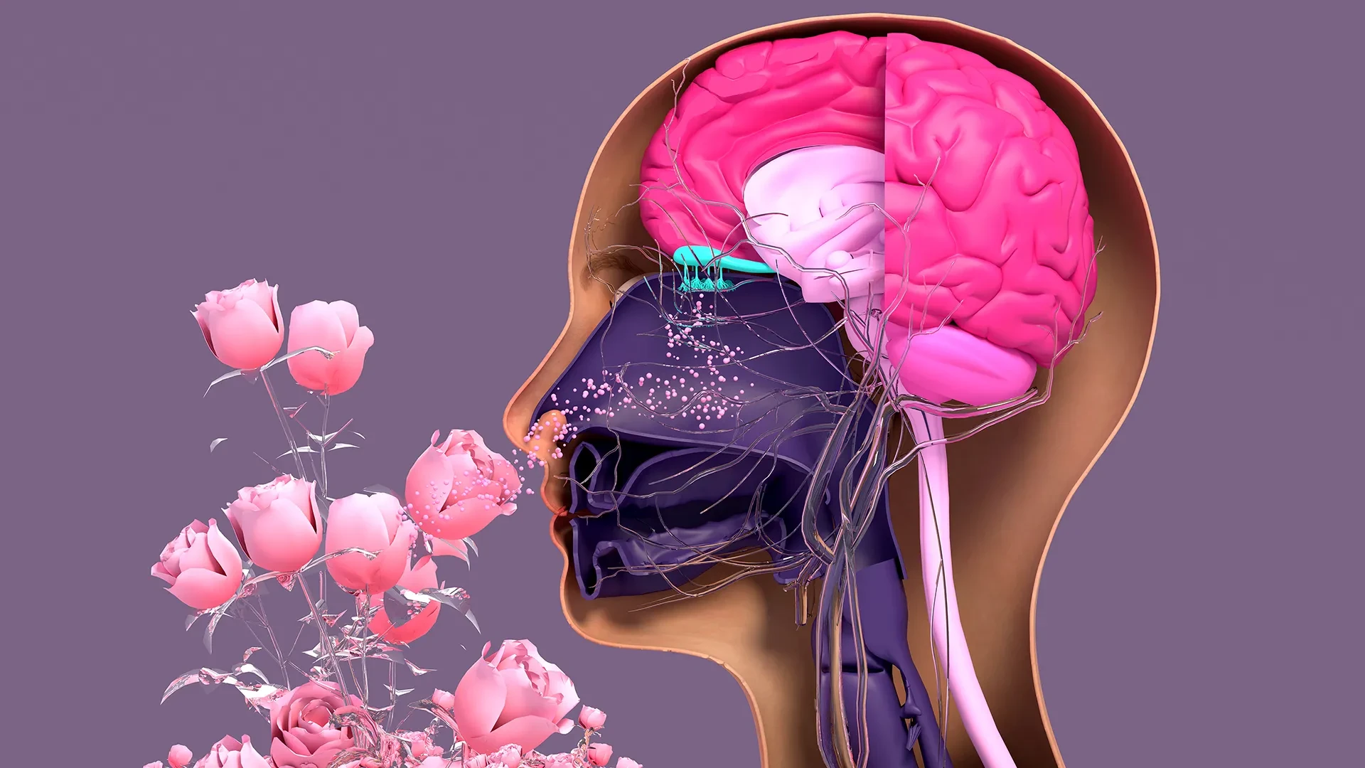
The human sense of smell is governed by sensory neurons located deep within the nasal cavity. Unlike the delicate neurons of the central nervous system, which are notoriously difficult to repair once damaged, these olfactory neurons possess a unique and robust capacity for regeneration. Throughout a person’s life, these cells are in a constant state of turnover, dying off and being replaced by new ones. This resilience is a biological necessity, as the nasal lining is one of the few places where the nervous system is directly and perpetually exposed to the external environment, including pathogens, pollutants, and physical trauma.
However, this regenerative capability is not invincible. Factors such as severe viral infections—most notably COVID-19—prolonged exposure to environmental toxins, and the natural process of aging can severely hamper the ability of these cells to replicate. When the regenerative cycle is disrupted, the result is often a partial loss of smell, known as hyposmia, or a total loss, known as anosmia. To better understand why this process fails and how it might be jump-started, the Tufts team created a new, accessible three-dimensional olfactory tissue model, or "organoid," using mouse cells. This model allows scientists to observe the birth of neurons in real-time and investigate the cellular breakdown that occurs during disease and senescence.
The findings, recently published in the journal Cell Reports Methods, provide a detailed look at the internal communication system of the nose. The research focuses on two primary types of stem cells found in the olfactory epithelium: horizontal basal cells (HBCs) and globose basal cells (GBCs). Traditionally, GBCs were viewed as the primary workhorses of regeneration, actively dividing to replace lost neurons, while HBCs were seen as a reserve population that remained quiet unless a catastrophic injury occurred. The new study challenges this binary view, suggesting a much more integrated relationship between the two cell types.
Brian Lin, a research assistant professor in the Department of Developmental, Molecular and Chemical Biology and the study’s senior author, notes that the research points toward a complex interdependency. According to Lin, the study suggests that these two stem cells may be fundamentally reliant on one another to function correctly. The HBCs, which were once thought to be passive bystanders in the day-to-day maintenance of the nasal tissue, may actually be providing essential support that allows for the production of new neurons and the successful repair of damaged tissue.
Using the 3D organoid model, the research team was able to isolate and identify a specific subpopulation of HBCs. These particular cells are characterized by their production of a protein known as KRT5. In the controlled environment of the organoid, the researchers observed that these KRT5-producing HBCs were not dormant at all; instead, they were actively facilitating the generation of new olfactory neurons.
To confirm the importance of these cells, the team conducted depletion experiments. When the KRT5-marked HBCs were selectively removed from the organoid cultures, the regenerative process stalled significantly. The generation of new neurons was severely impaired, proving that these "dormant" cells are, in fact, essential players in the biological theater of the nose. This discovery opens new avenues for therapeutic research, as it suggests that targeting HBCs might be a viable way to treat long-term smell loss in patients where the GBC population has been exhausted or damaged.
Beyond the basic mechanics of regeneration, the study also sought to address the common clinical observation that the sense of smell fades with age. The researchers utilized cells harvested from mice of varying ages to grow organoids, comparing the regenerative potential of young tissue against that of older specimens. The results were telling: there was a marked decline in the ability of cells from older mice to produce new sensory neurons.
Lin explains that this decline appears to be linked to changes in the stem cell populations over time. The team believes that as an organism ages, the population of globose basal cells (GBCs) begins to dwindle, or their ability to divide becomes compromised. While the HBCs may still be present, the loss of their cellular partners—the GBCs—disrupts the "interdependent" cycle of growth. Lin emphasizes that more work is required to fully test this hypothesis, but the ultimate goal is to find ways to rejuvenate these aging cell populations, potentially restoring sensory function to the elderly.
An Easy-To-Use Model
One of the most significant contributions of this study is the nature of the model itself. The development of the 3D organoid was led by Juliana Gutschow Gameiro, a former Ph.D. student who visited the Graduate School of Biomedical Sciences at Tufts from the State University of Londrina in Brazil. From the outset, Gameiro and Lin were committed to creating a research tool that was not only effective but also accessible to the wider scientific community.
In the world of high-level biological research, many advanced models require exorbitant funding, specialized equipment, and highly specific expertise in stem cell biology. Gameiro’s model was designed to bypass these barriers. By creating an organoid that is relatively easy to produce and maintain, the team has provided a blueprint that can be used by labs with limited resources.
This accessibility is particularly timely. In the wake of the global pandemic, the study of the olfactory system has moved from a niche interest to a major focus of medical research. The prevalence of "long COVID" symptoms, which frequently include a persistent lack of taste and smell, has drawn researchers from diverse fields—such as neurology, immunology, and gerontology—into the study of olfactory epithelial cells.
Lin highlights the importance of this cross-disciplinary interest, noting that the team wanted to develop a tool that non-stem cell biologists could easily adopt. By lowering the barrier to entry, they hope to accelerate the global understanding of how olfactory neurons regenerate and, conversely, what specific triggers cause that process to diminish or fail entirely in the face of disease.
Next Step: A Human Organoid
While the mouse-tissue model has provided groundbreaking insights, it serves as a stepping stone toward a more ambitious goal: the creation of a human olfactory organoid. Such a model would represent a "holy grail" for pharmacological research in this field, allowing scientists to screen potential drugs and therapies on living human tissue without the ethical and logistical complications of full-scale human trials.
The advantages of organoids over traditional research methods are substantial. Currently, many researchers rely on whole animal models, which may not perfectly replicate human biology, or existing human cell cultures, which are often two-dimensional and fail to capture the complex architecture of real organs. Organoids bridge this gap, offering a "mini-organ" that functions much like the real thing. They have already revolutionized the study of the lungs, kidneys, and brain, but the human olfactory system has remained a challenge.
The primary obstacle to creating a human olfactory organoid is the difficulty of obtaining pure tissue samples. As Lin explains, the process of collecting these cells from a living person is invasive and technically difficult. It involves anesthetizing the individual and using a specialized brush—similar to the long swabs used in COVID-19 testing—to reach deep into the uppermost reaches of the nasal cavity.
Even when successful, this collection process yields a mixture of different cell types. In the nose, respiratory stem cells (responsible for the lining that produces mucus) and olfactory stem cells (responsible for smell) live in close proximity. Unlike the mouse model, where these populations can be separated with relative ease, human samples are often a tangled mix that is difficult to sort.
The Tufts research team is now shifting its focus toward overcoming this hurdle. Their next challenge involves developing a simple, cost-effective technique to separate human olfactory stem cells from their respiratory counterparts. Once isolated, the goal is to "coax" these cells to grow and organize into 3D structures in the lab.
If successful, these human organoids could provide the first reliable platform for testing treatments for anosmia. Whether the cause is a viral infection, a neurodegenerative condition like Parkinson’s disease—where smell loss is often an early warning sign—or simply the passage of time, the ability to observe human nasal regeneration in a dish could lead to the first effective clinical interventions for a sense that is too often taken for granted until it is gone.