
In a significant advancement for sensory biology and regenerative medicine, researchers at Tufts University School of Medicine and the Graduate School of Biomedical Sciences (GSBS) have pioneered a new three-dimensional model to study how the human nose maintains its sense of smell. The study, conducted in collaboration with international colleagues, has led to a surprising discovery: a type of stem cell long considered to be largely dormant may actually be a primary driver in the preservation and repair of olfactory tissue.
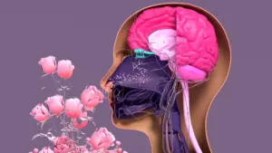
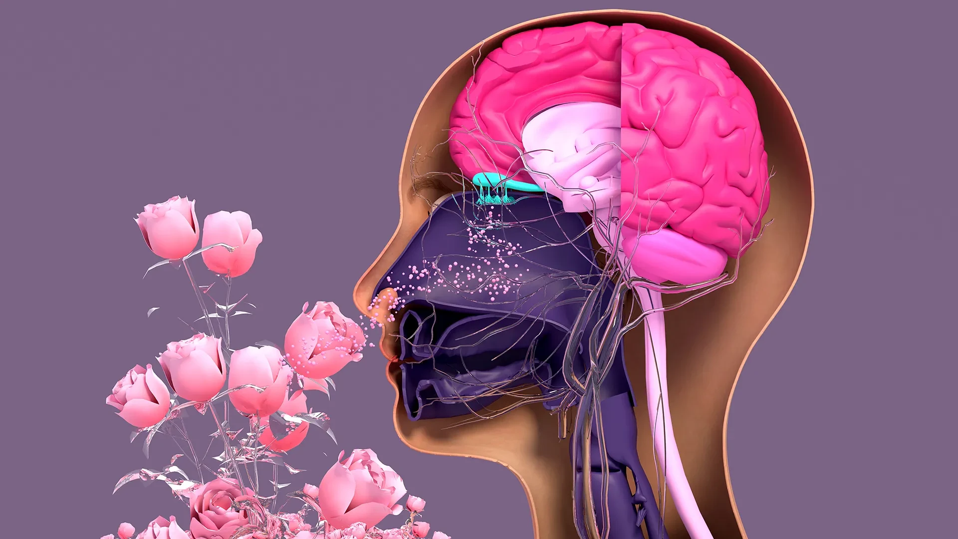
The research focuses on the olfactory epithelium—the specialized tissue inside the nasal cavity responsible for detecting odors. Unlike the delicate cells of the central nervous system, such as those in the brain or spinal cord, the sensory neurons in the nose possess a rare and remarkable capacity for regeneration. Throughout a person’s life, these neurons are constantly being replaced, a biological necessity given their position on the body’s front lines, where they are perpetually exposed to environmental hazards, including pollutants, toxins, and pathogens.
However, this regenerative engine is not invincible. Factors ranging from the natural process of aging to severe viral infections—most notably COVID-19—can interrupt the cycle of renewal. When the production of new neurons slows or ceases, the result is often a partial loss of smell (hyposmia) or a total loss (anosmia), conditions that can profoundly impact quality of life, safety, and even mental health. To better understand these failures and seek potential treatments, the Tufts team developed a sophisticated yet accessible 3D "organoid" model of mouse olfactory tissue.
The Interplay of Stem Cells: HBCs and GBCs
The study, recently published in the journal Cell Reports Methods, provides an unprecedented look at the cellular "cross-talk" required to keep the sense of smell functioning. The researchers focused on two primary populations of stem cells found in the nasal lining: horizontal basal cells (HBCs) and globose basal cells (GBCs).
Historically, the scientific community viewed these two cell types through a relatively narrow lens. Globose basal cells were recognized as the "workhorses" of the nose—active, rapidly dividing cells that directly give rise to new sensory neurons. In contrast, horizontal basal cells were largely characterized as a reserve population. It was believed that HBCs remained in a state of dormancy, or "quiescence," only springing into action in the event of catastrophic tissue damage.
The Tufts study challenges this traditional hierarchy. Using their new 3D organoid model, the team discovered that these two stem cell populations are far more interdependent than previously understood.
"Our research suggests that these two stem cells may be interdependent," explains Brian Lin, the study’s senior author and a research assistant professor in the Department of Developmental, Molecular and Chemical Biology at Tufts. "One type that we thought was largely dormant—HBCs—may actually play a crucial role in supporting the production of new neurons and the repair of damaged tissue."
By observing the cells within the 3D architecture of the organoid, the researchers identified a specific subpopulation of HBCs that produce a protein known as KRT5. These KRT5-positive cells appeared to be the primary coordinators of the regenerative process. In a series of controlled experiments, the team selectively depleted these specific HBCs from the organoid cultures. The results were immediate and clear: without the support of these "dormant" cells, the generation of new olfactory neurons was significantly impaired. This suggests that HBCs are not merely a backup system for emergencies but are essential players in the day-to-day maintenance of the olfactory system.
Insights into Aging and Sensory Decline
One of the most pressing questions in olfactory research is why the sense of smell inevitably declines as people grow older. This loss is more than a minor inconvenience; it is often linked to decreased appetite, depression, and in some cases, it serves as an early warning sign for neurodegenerative diseases like Alzheimer’s and Parkinson’s.
To investigate this, Lin and his colleagues utilized their model to compare cells harvested from mice of various ages. When they attempted to grow organoids using cells from older subjects, they observed a marked decline in the ability to generate new sensory neurons.
"We found a decline in the ability of the older mice cells to generate new neurons," says Lin. "We think this is due to a decrease in the GBC population as we age, but we need to do more work to test this hypothesis and if so, develop ways to rejuvenate them."
The 3D model allows researchers to isolate these variables in a way that traditional 2D cell cultures or whole-animal studies cannot. By observing how the interaction between HBCs and GBCs falters over time, the team hopes to identify the molecular "brakes" that slow down regeneration in the elderly. If the GBC population is indeed the bottleneck, future therapies might focus on stimulating these cells or enhancing the supportive signals they receive from the HBCs to restore youthful regenerative capacity.
An Easy-To-Use Model for Global Research
The development of the organoid itself is as much a part of the breakthrough as the biological findings it produced. The study’s lead author, Juliana Gutschow Gameiro, a former Ph.D. student who visited the Graduate School of Biomedical Sciences at Tufts from the State University of Londrina in Brazil, was instrumental in this effort.
Gutschow Gameiro and Lin set out with a specific goal: to create a model that was not only effective but also accessible. In the world of high-tech biological research, many advanced models require prohibitively expensive equipment, proprietary reagents, or highly specialized training that limits their use to the world’s most well-funded institutions.
"Because loss of smell is associated with COVID-19, as well as with Parkinson’s disease and other conditions, a much larger number of researchers from a variety of different fields have begun researching olfactory epithelial cells in the last few years," says Lin.
Recognizing this surge in interest, the team designed the mouse-tissue organoid to be "easy-to-create." By providing a simplified protocol that can be replicated in labs with limited funds and standard equipment, they hope to democratize the study of the sense of smell. This approach allows scientists who may not be stem cell specialists—such as virologists studying how COVID-19 enters the nervous system or toxicologists looking at air pollution—to incorporate high-quality olfactory tissue models into their work.
Next Step: A Human Organoid
While the mouse model has provided a wealth of new information regarding the fundamental mechanics of stem cell interdependence, the ultimate objective of the Tufts team is to translate these findings into human health applications. The next major milestone on their roadmap is the development of a human olfactory organoid.
The potential for a human-specific model is vast. In the pharmaceutical industry, organoids serve as a bridge between early-stage laboratory research and human clinical trials. They allow for "pre-clinical" drug screening that is often more accurate than animal testing, as they utilize actual human cells and replicate the complex 3D structure of human organs. While organoids for the lungs, kidneys, and liver have been in use for several years, the olfactory system has remained a difficult frontier.
The primary obstacle is the difficulty of obtaining pure, viable human olfactory tissue. "It’s challenging to get pure olfactory tissue from humans," Lin notes. Currently, the process involves a procedure where a patient is anesthetized, and a medical professional uses a specialized brush—similar to a long COVID-19 testing swab—to reach deep into the upper nasal cavity to collect cells.
However, the biology of the human nose presents a complication that the mouse model does not: the close proximity and intermingling of different tissue types. When cells are collected via the brush method, the sample typically contains a mixture of respiratory stem cells (which line the majority of the nasal passage) and olfactory stem cells. In the laboratory, these two types are notoriously difficult to separate, making it hard to grow a pure olfactory organoid that accurately reflects the sensory system.
The research team is now focused on overcoming this technical hurdle. Their goal is to develop a simple and cost-effective technique to isolate human olfactory stem cells from respiratory ones and "coax" them into forming the same supportive 3D structures seen in their mouse model.
Once perfected, a human olfactory organoid could revolutionize how we treat sensory loss. It would provide a platform to test new drugs that could "wake up" dormant stem cells or replace those lost to disease. For the millions of people worldwide suffering from long-term smell loss following viral infections or due to the progression of age, the work being done in the labs at Tufts offers a promising pathway toward reclaiming one of the most fundamental ways we experience the world.