The human body possesses an almost miraculous capacity for self-repair. When the skin is sliced or the epithelial linings of internal organs are damaged by trauma or disease, a complex biological symphony begins, working to restore the integrity of the tissue. For decades, scientists have observed this phenomenon, known as compensatory proliferation, where a burst of cellular growth replaces lost or damaged tissue. Yet, despite its fundamental importance to survival, the precise molecular triggers that allow a wounded organ to "know" how to rebuild itself have remained one of biology’s most enduring mysteries.
Now, a groundbreaking study from the Weizmann Institute of Science has pulled back the curtain on this process, revealing a surprising and paradoxical mechanism. According to research published in the prestigious journal Nature Communications, the very enzymes responsible for cellular "suicide" are also the key architects of survival and regeneration. While this discovery offers profound insights into how healthy tissues heal, it also provides a sobering explanation for one of the most frustrating challenges in modern medicine: why certain cancers return with increased aggression and resistance after being targeted by radiation and chemotherapy.
The research, led by Prof. Eli Arama and Dr. Tslil Braun of Weizmann’s Molecular Genetics Department, suggests that a specific population of cells can initiate the process of programmed death only to halt it midway, effectively "daring" to survive. These survivors then become the primary drivers of tissue regrowth, but they also pass on an enhanced resistance to their descendants—a trait that, in a cancerous context, can lead to treatment-resistant tumors.
A Half-Century Mystery: The Roots of Compensatory Proliferation
The scientific community’s fascination with compensatory proliferation dates back to the 1970s. At the time, researchers working with Drosophila melanogaster—the common fruit fly—made a startling observation. When fly larvae were exposed to high doses of ionizing radiation, the epithelial tissues that would eventually form the adult wings suffered catastrophic damage. By all accounts, the larvae should have developed into adults with severely deformed or missing wings.
Instead, the larvae demonstrated an incredible resilience. They managed to regenerate the damaged areas so effectively that the resulting adult flies possessed fully functional, perfectly formed wings. This indicated that the remaining healthy cells were receiving a signal to multiply rapidly and fill the void left by their deceased neighbors.
While this phenomenon was later observed across various species, including mammals and humans, the "how" remained elusive. What was the signal? And which specific cells were responsible for leading the charge? For fifty years, the prevailing theory was that dying cells released "stress signals" that prompted their healthy neighbors to divide. However, the Weizmann team suspected the story was far more complex, involving a specialized group of cells that occupy a middle ground between life and death.
When Cell Death Machinery Promotes Survival
To understand the Weizmann discovery, one must first look at apoptosis, the process of programmed cell death. Apoptosis is a vital "housekeeping" function of the body, used to eliminate cells that are no longer needed, are aging, or have suffered DNA damage that could lead to cancer. It is a tidy, controlled process that prevents the leakage of harmful cellular contents into surrounding tissue.
The primary executioners of apoptosis are a family of enzymes called caspases. The process generally follows a strict hierarchy: an "initiator" caspase is activated by a stress signal, which then triggers "effector" or "executioner" caspases. These executioners act like molecular scissors, shredding the cell’s internal proteins and ensuring its demise.
However, the scientific consensus on caspases has shifted over the last twenty years. Prof. Eli Arama was among the early pioneers who suggested that these "death enzymes" might have non-lethal roles. His previous work demonstrated that caspases are involved in various essential life processes, such as the shaping of sperm cells and the development of certain tissues, where they prune cellular components without killing the entire cell.
In this new study, Arama and his team hypothesized that these non-lethal caspase functions might be the "missing link" in compensatory proliferation. They suspected that some cells might activate the death machinery but use it as a signaling tool rather than a self-destruct button.
Finding Cells That Start To Die but Survive
To test this hypothesis, Dr. Tslil Braun recreated the classic 1970s radiation experiments, but with a sophisticated modern twist. Using advanced genetic engineering, the team created a "delayed sensor" system in fruit fly larvae. This sensor allowed the researchers to track the history of individual cells with extreme precision.
Specifically, the sensor was designed to flag any cell that had activated its initiator caspase but had not actually died. This allowed the team to distinguish between cells that were completely healthy, cells that had died, and a mysterious third group: the survivors of a near-death experience.
"We set out to identify cells that push the self-destruct button but survive anyway," Dr. Braun explained. "This is how we discovered a population of cells we named DARE cells."
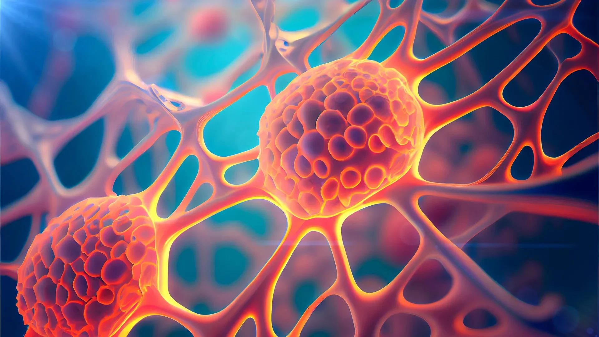
The acronym DARE stands for "Death-Associated Resistant Entities." These cells were a revelation. Despite being hit with radiation levels that killed many of their neighbors, DARE cells activated their initiator caspases and then simply refused to die. More importantly, they became hyper-active. Within 48 hours of the radiation exposure, these DARE cells multiplied so rapidly that they replenished nearly half of the damaged tissue.
However, the DARE cells were not acting alone. The researchers identified a second population of death-resistant cells they called NARE cells (Non-apoptotic Resistant Entities). Unlike the DARE cells, the NARE cells never activated their initiator caspases at all. While both populations contributed to the final regenerated tissue, the DARE cells were the essential catalysts.
"When we removed DARE cells from the system, compensatory proliferation disappeared entirely," Braun noted. The study found that DARE cells were effectively the "first responders" of the regeneration process, activated by signals from their dying neighbors and then coordinating the repair efforts with the NARE cells.
How DARE Cells Escape Their Death Sentence
The discovery of DARE cells led to a critical question: how do these cells stop the "executioner" caspases from finishing the job once the "initiator" has already been activated?
The Weizmann team found that the answer lies in the cell’s internal architecture. In a typical apoptotic event, the initiator caspase moves freely to activate the executioners. In DARE cells, however, the process is physically blocked. The researchers identified a molecular motor protein that tethers the initiator caspase to the cell’s outer membrane. By "pinning" the enzyme to the membrane, the motor protein prevents it from reaching and activating the executioner caspases in the rest of the cell.
"We observed that although the initiator caspase is activated in these cells, the cellular death process stops there and does not progress to the next stage," Arama explains.
To prove this, the researchers silenced the gene responsible for this motor protein. Without the tether, the DARE cells were no longer able to stop the death process; they proceeded to die just like their neighbors, and the tissue’s ability to regenerate was severely impaired. Conversely, the researchers noted that overactivation of this same motor protein has been previously observed in various human cancers, suggesting that tumors may be hijacking this natural healing mechanism to stay alive.
Surviving Radiation Can Make Cells Harder To Kill
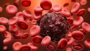
The implications for oncology are significant. One of the most harrowing aspects of cancer treatment is the recurrence of a tumor. Often, a patient will undergo radiation therapy, and the tumor will shrink or disappear, only to return months or years later. When it returns, it is frequently more aggressive and completely resistant to the treatments that worked the first time.
The Weizmann study provides a biological explanation for this "acquired resistance." The researchers found that the DARE cells don’t just survive radiation; they evolve because of it.
"We wanted to understand whether resistance to death is inherited by the descendants of death-resistant cells," Arama said. The results were startling. When the researchers irradiated the regenerated tissue a second time, they found that the descendants of the original DARE cells were seven times more resistant to cell death than the cells in the original, untreated tissue.
This suggests that the process of surviving the first round of radiation "trains" the cells and their offspring to be virtually unkillable. In a healthy body, this creates a robust, damage-resistant patch of skin or organ lining. In a tumor, however, this creates a reservoir of "super-cells" that can withstand subsequent rounds of therapy, leading to the aggressive recurrences seen in clinical settings.
A Feedback Loop Keeps Regeneration Under Control
If DARE cells are so efficient at multiplying and resisting death, what stops them from growing forever? In a healthy system, regeneration must be finite; it must stop once the tissue is restored. If it doesn’t, the result is the uncontrolled growth characteristic of a tumor.
In the final stage of their study, the researchers uncovered a elegant signaling system that maintains this balance. They discovered a negative-feedback loop between the DARE and NARE cell populations.
DARE cells, the primary survivors, secrete growth signals that encourage the nearby NARE cells to multiply and help fill the gap. In return, as the NARE cells grow and the tissue reaches its proper density, the NARE cells begin secreting their own signals that tell the DARE cells to stop dividing. This cross-talk ensures that the "repair crew" knows exactly when the job is finished, preventing the overgrowth that could lead to a secondary health crisis.
From Tissue Repair to Cancer Treatment
While the study was conducted using fruit fly models, the researchers are optimistic about its translation to human medicine. Historically, many of the most fundamental discoveries in genetics—from the way embryos develop to the way genes are turned on and off—were first discovered in fruit flies before being confirmed in humans.
"We hope that the knowledge gained here can be translated into an understanding of the mechanisms that balance growth and confer resistance to cell death in human tissues," Arama concludes.
The discovery offers two potential paths for future medical breakthroughs. On one hand, by understanding how DARE cells work, doctors might be able to develop therapies that "boost" this mechanism to help patients recover from severe burns, chronic wounds, or organ damage.
On the other hand, in the fight against cancer, this research provides a new target. If scientists can develop drugs that inhibit the "tethering" motor protein or disrupt the DARE cells’ survival signals, they may be able to prevent cancer cells from surviving the initial round of radiation. By forcing these cells to complete the apoptosis process they started, doctors could potentially eliminate the "seeds" of future, more resistant tumors.
The findings from the Weizmann Institute highlight the dual nature of our biological systems: the very pathways that allow us to heal from injury are the same ones that can be exploited by disease. Understanding this delicate balance between life and death is the next great frontier in both regenerative medicine and oncology.