

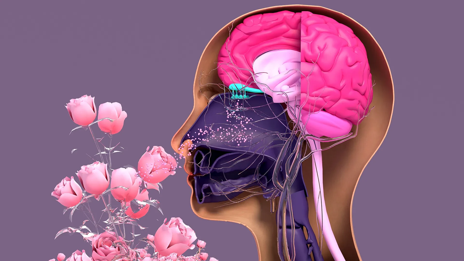
In a significant advancement for the field of sensory biology, researchers at Tufts University School of Medicine and the Graduate School of Biomedical Sciences (GSBS), along with an international team of colleagues, have unveiled a sophisticated three-dimensional model designed to study the complex regeneration of nerve tissue within the human nose. The study, which utilizes this newly devised "organoid" system, has led to a surprising discovery regarding the cellular architecture of our olfactory system: a type of stem cell long dismissed as largely dormant may actually be a primary driver in maintaining our ability to smell.
The findings, published in the journal Cell Reports Methods, offer a fresh perspective on how the body repairs itself after injury or illness and why these regenerative powers often fail as we age. By providing a more accessible and accurate way to observe the birth of new neurons, the research team hopes to pave the way for future treatments for millions of people suffering from olfactory dysfunction—a condition that has gained global attention in the wake of the COVID-19 pandemic.
The Unique Resilience of the Olfactory System
Unlike the majority of cells found within the central nervous system, which are notoriously difficult to repair or replace once damaged, the sensory neurons located in the nasal cavity possess a remarkable, lifelong capacity for regeneration. These neurons are uniquely positioned; they are the only part of the nervous system directly exposed to the external environment. Every breath we take introduces a barrage of potential threats, including viral pathogens, environmental toxins, pollutants, and physical irritants.
To survive this constant environmental assault, the body has evolved a highly efficient system for "recycling" and replacing these neurons. However, this process is not infallible. Significant viral infections, such as COVID-19 or the flu, prolonged exposure to industrial chemicals, and the natural biological wear and tear of aging can all disrupt this delicate cycle. When the ability of these cells to replicate is diminished or halted, the result is hyposmia (a partial loss of smell) or anosmia (a complete loss of smell).
Understanding why this process succeeds in some scenarios and fails in others has been a primary challenge for scientists. To address this, the Tufts team sought to create a laboratory model that could mimic the complex environment of the nasal cavity more effectively than traditional two-dimensional cell cultures.
The Role of Horizontal Basal Cells: From Dormancy to Action
At the heart of the team’s research is a deep dive into the two primary types of stem cells found in the olfactory epithelium: horizontal basal cells (HBCs) and globose basal cells (GBCs). Historically, the scientific community has viewed GBCs as the primary workhorses of the nose—the active, rapidly dividing cells responsible for the day-to-day replacement of sensory neurons. In contrast, HBCs were largely characterized as "reserve" cells—dormant entities that only sprung into action under conditions of extreme tissue trauma.
However, the new 3D mouse model developed at Tufts has challenged this long-standing hierarchy. The researchers discovered that these two cell types are far more interdependent than previously understood.
"Our research suggests that these two stem cells may be interdependent," explained Brian Lin, the study’s senior author and a research assistant professor in the Department of Developmental, Molecular and Chemical Biology at Tufts. "One type that we thought was largely dormant—HBCs—may actually play a crucial role in supporting the production of new neurons and the repair of damaged tissue."
By observing the cells within the organoid model, the team identified a specific subpopulation of HBCs. These particular cells are distinguished by their production of a protein known as KRT5 (Keratin 5). The study found that these KRT5-producing HBCs are not merely sitting in reserve; they are active participants that support the generation of new olfactory neurons.
The evidence for this role was clear: when the researchers selectively removed these specific HBCs from the organoid cultures, the production of new neurons was significantly impaired. This suggests that HBCs provide a necessary "niche" or support system that allows the GBCs to function correctly. Without the influence of the HBCs, the regenerative cycle essentially stalls.
Aging and the Decline of Sensory Regeneration
The study also shed light on one of the most common causes of smell loss: the natural process of aging. It is a well-documented medical fact that the sense of smell often dulls as people enter their later decades, but the cellular mechanism behind this decline has remained somewhat elusive.
To investigate this, the Tufts team utilized their model to compare cells harvested from mice of various ages. The results showed a marked difference in regenerative capacity. Cells taken from older mice were significantly less effective at generating new neurons within the 3D organoid environment.
"We found a decline in the ability of the older mice cells to generate new neurons," Lin noted. "We think this is due to a decrease in the GBC population as we age, but we need to do more work to test this hypothesis and if so, develop ways to rejuvenate them."
This finding is particularly relevant for the development of geriatric medicine. If the decline in smell is tied specifically to the depletion of globose basal cells or the failure of the horizontal basal cells to support them, it opens the door for targeted therapies that could potentially "restart" the regenerative process in older adults.
An Easy-To-Use Model
One of the most significant contributions of this research is the nature of the model itself. In the world of high-level molecular biology, research can often be prohibitively expensive, requiring specialized equipment and massive budgets. However, the lead author of the study, Juliana Gutschow Gameiro, was determined to create a tool that was accessible to the wider scientific community.
Gameiro, a former Ph.D. student who visited the Graduate School of Biomedical Sciences at Tufts from the State University of Londrina in Brazil, focused on developing a 3D olfactory tissue model—or organoid—that was "easy-to-create."
"Because loss of smell is associated with COVID-19, as well as with Parkinson’s disease and other conditions, a much larger number of researchers from a variety of different fields have begun researching olfactory epithelial cells in the last few years," said Lin.
The surge in interest following the pandemic meant that scientists who were not necessarily experts in stem cell biology were suddenly tasked with understanding the nasal environment. By making the model simple and inexpensive, the Tufts team has provided a platform for labs with limited resources to conduct high-quality research. This democratization of the technology allows for a more diverse range of studies into how neurons are formed and what causes the process to fail in the face of disease.
Next Step: A Human Organoid
While the current success involves a mouse-tissue model, the researchers have their sights set on a much larger goal: the creation of a human olfactory organoid. Such a model would represent a massive leap forward for clinical medicine, acting as a bridge between laboratory research and patient care.
In the modern drug development pipeline, organoids are becoming increasingly vital. They allow for pre-clinical trials that are faster, cheaper, and often more accurate than animal testing. Because an organoid is grown from actual tissue, it can provide a realistic preview of how a human organ might respond to a specific drug or treatment. While organoids have already been successfully developed for the lungs, kidneys, and liver, the human olfactory system has proven to be a much tougher nut to crack.
The primary obstacle is the difficulty of obtaining pure, uncontaminated human olfactory tissue. Unlike mice, where tissue can be harvested with precision, collecting samples from humans is an invasive and complicated process. It typically involves anesthetizing the patient and using a specialized brush—similar to the swabs used for COVID-19 tests but pushed much deeper into the nasal cavity—to scrape the upper reaches of the nose.
The challenge, according to Lin, is that this process inevitably collects a mixture of different cell types. "It’s challenging to get pure olfactory tissue from humans," he explained. In the resulting sample, human respiratory stem cells are often mixed with olfactory stem cells, and currently, they are extremely difficult to separate.
The research team’s next major hurdle is to refine their techniques to isolate these human olfactory stem cells. Once they can separate these cells from the surrounding respiratory tissue, they can begin coaxing them to grow into 3D organoids in the lab.
The ultimate vision is a future where a patient suffering from chronic smell loss—whether from long-COVID, neurodegenerative disease, or age—could have their own cells used to grow a personalized organoid. Doctors could then use that organoid to screen various drugs and regenerative therapies to see which one works best for that specific individual before ever starting a clinical trial.
By uncovering the "hidden" power of horizontal basal cells and providing a blueprint for accessible 3D modeling, the Tufts researchers have not only deepened our understanding of the nose but have also provided a new sense of hope for restoring one of our most vital connections to the world around us.