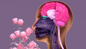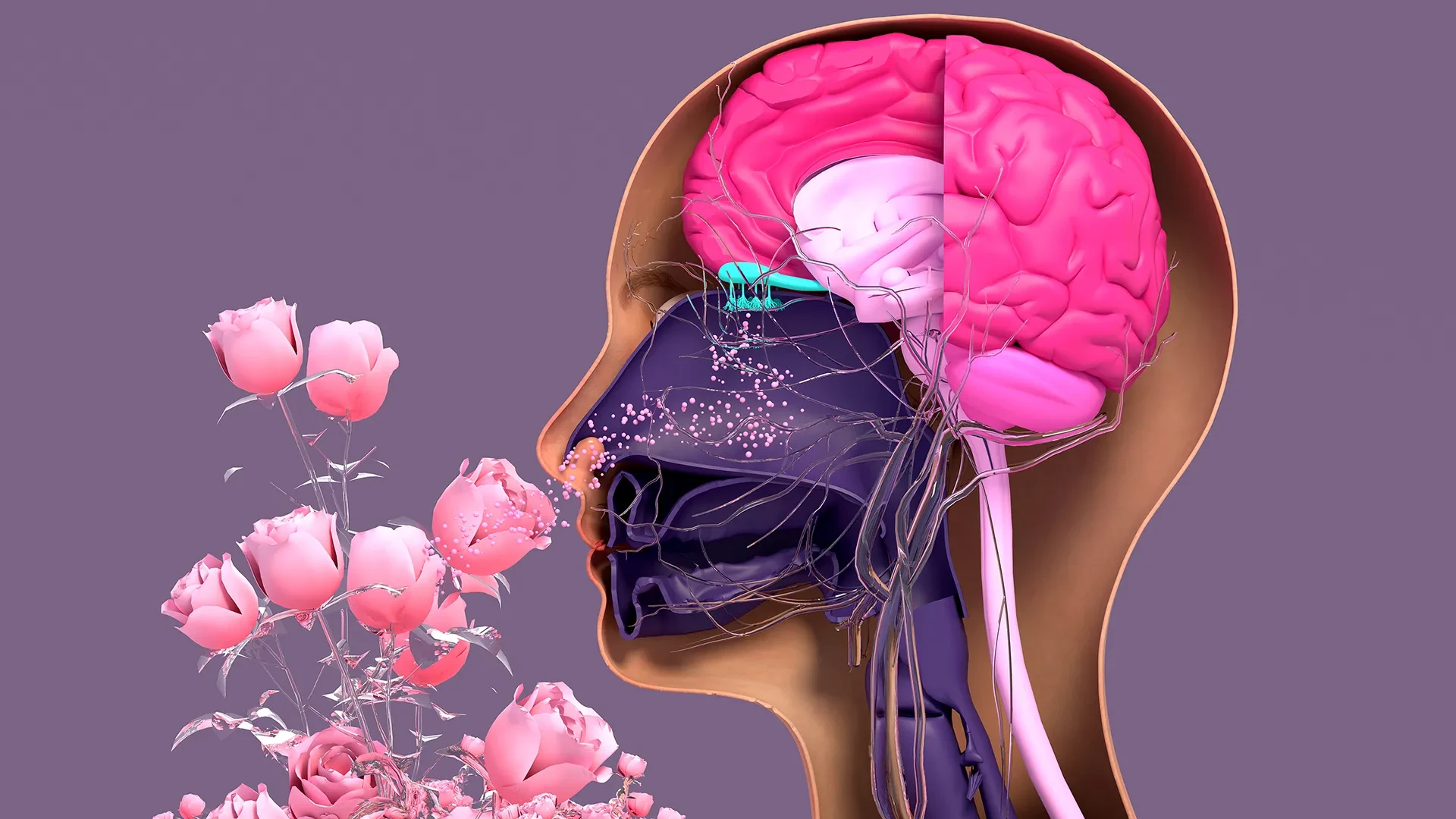
In the complex architecture of the human sensory system, the sense of smell often occupies a paradoxical position. It is perhaps our most primal sense, capable of triggering vivid memories or alerting us to invisible dangers, yet it remains one of the least understood in terms of its regenerative capacity and the mechanisms of its decline. While the neurons of the central nervous system are notoriously limited in their ability to repair or replace themselves, the sensory neurons located within the nasal cavity are remarkably resilient. They possess a unique, lifelong ability to regenerate, a necessary adaptation given their constant exposure to an environment filled with pollutants, allergens, and pathogens.
However, this resilience is not infinite. Factors ranging from viral infections, such as the global impact of COVID-19, to toxic environmental exposures and the natural progression of aging can severely hinder the body’s ability to maintain this delicate sensory interface. When the regenerative cycle fails, the result is often a partial or total loss of smell—a condition that can significantly diminish quality of life.
In a significant step forward for the field of regenerative medicine, a team of researchers at Tufts University School of Medicine and the Graduate School of Biomedical Sciences (GSBS), along with their international colleagues, has developed a sophisticated new tool to study this process. By creating a three-dimensional organoid model of olfactory tissue, the team has challenged long-standing assumptions about the "dormant" nature of certain stem cells in the nose, revealing that they play a far more active and essential role in preserving our sense of smell than previously recognized.
A New Perspective on Nasal Stem Cells
The research, recently published in the journal Cell Reports Methods, focuses on the intricate dance between two primary types of stem cells found in the olfactory epithelium: horizontal basal cells (HBCs) and globose basal cells (GBCs). Traditionally, the scientific community viewed GBCs as the primary "workhorses" of nasal regeneration—the active progenitors that divide frequently to replace aging or damaged neurons. In contrast, HBCs were often characterized as a reserve population, a "dormant" backup system that only sprung into action in the event of catastrophic tissue damage.
The Tufts study, led by senior author Brian Lin, a research assistant professor in the Department of Developmental, Molecular and Chemical Biology, suggests this binary view is incomplete. Using their newly devised 3D mouse model, the researchers discovered that these two cell types exist in a state of interdependence.
"Our research suggests that these two stem cells may be interdependent," Lin explained. "One type that we thought was largely dormant—HBCs—may actually play a crucial role in supporting the production of new neurons and the repair of damaged tissue."
This discovery shifts the narrative of nasal regeneration from a simple replacement process to a complex, collaborative effort. The HBCs appear to provide a necessary support structure or signaling environment that allows the GBCs to function effectively. Without the active participation of the HBCs, the entire assembly line of new sensory neurons begins to stall.
The Power of the 3D Organoid Model
To reach these conclusions, the team had to overcome the limitations of traditional laboratory methods. In many previous studies, researchers relied on two-dimensional cell cultures, which fail to capture the physical and chemical complexities of living tissue. To bridge this gap, the Tufts team developed a three-dimensional olfactory organoid—a "mini-organ" grown in a dish that mimics the structure and function of the mouse nasal lining.