The transmission cycle begins when parasite eggs are shed into the environment through the urine or feces of infected mammalian hosts, which include humans as well as various domestic and wild animals. In rural agricultural settings, particularly where sanitation infrastructure may be limited, these excreted eggs frequently contaminate nearby freshwater bodies, drainage ditches, and flooded agricultural fields.
When the eggs land in suitable freshwater conditions, they hatch into a free-swimming, microscopic larval form. These aquatic larvae do not immediately infect human hosts; instead, they are biological obligates that must first locate specific species of freshwater snails that serve as intermediate hosts. Upon identifying a suitable snail, the larva actively bores through the mollusk’s outer tissue to gain entry. Inside the soft tissues of the snail, the parasite undergoes two distinct developmental life stages, reproducing asexually and multiplying significantly in overall number.
After completing this essential phase of development within the snail, late-stage larvae burst out of the intermediate host and re-enter the surrounding water column. At this stage, the larvae are known as cercariae. Physical characteristics of cercariae include a distinctive forked tail that aids in their propulsion through the water as they swim in search of a definitive mammalian host.
Unlike many other waterborne parasites that must be ingested to cause infection, cercariae possess the extraordinary biological capability to tunnel directly through intact host skin upon contact. This mode of transmission makes individuals who spend prolonged periods standing or working in freshwater particularly vulnerable to infection.
For a rice farmer in the Philippines, daily agricultural duties often involve wading barefoot or in thin clothing through flooded rice paddies. When cercariae present in the irrigation water encounter exposed human skin, they attach to the surface, shed their forked tails, and utilize specialized chemical secretions to penetrate the outer layers of the skin.
Once the larvae successfully penetrate the skin, they enter the host’s vascular system, initiating a complex migration through the cardiovascular network. The young parasites travel through the venous blood vessels to the right atrium and ventricle of the heart, pass through the capillary beds of the lungs, loop back through the left side of the heart, and are swept into the systemic arterial circulation.
Eventually, they navigate into the portal venous system of the liver. It is within the blood vessels of the liver that the immature flukes feed on host blood components, mature, and differentiate into adult male and female worms.
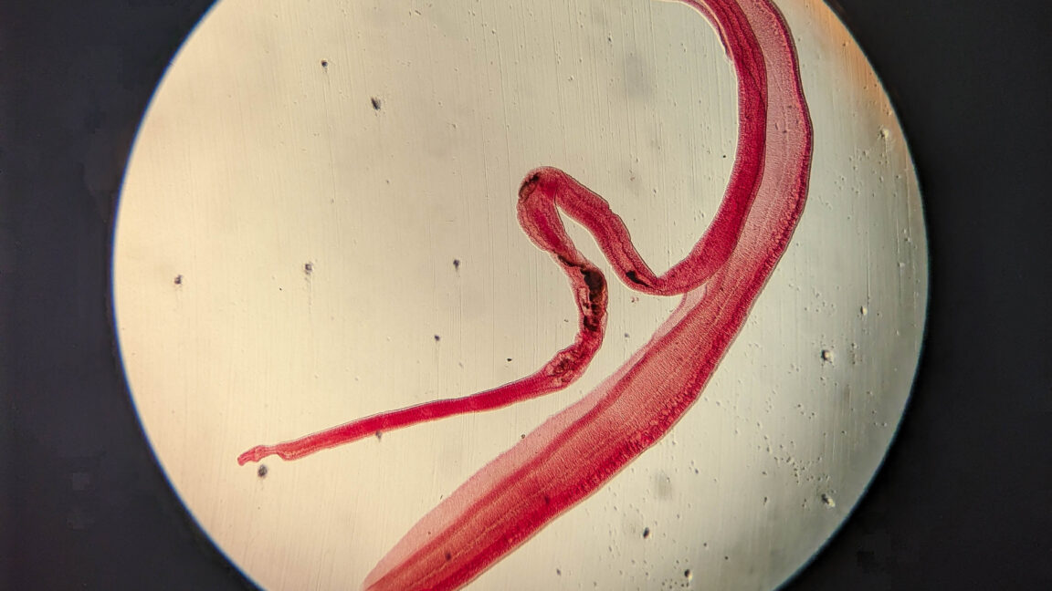
The structural anatomy and behavioral biology of mature Schistosoma worms are notably unique among parasitic flatworms. Adult male and female flukes live permanently paired together in a continuous biological embrace. The thicker adult male parasite features a deep, longitudinal groove along its body known as the gynaecophoric canal. The slender, elongated female worm tucks herself snugly inside this groove, where the coupled pair remains joined throughout their adult lives.