It began without any of the typical warning signs that usually precede a major neurological event. A woman working as a rice farmer in the Philippines was going about her day feeling entirely healthy—she had experienced no headache, no cognitive confusion, and no generalized body weakness. Then, completely out of nowhere, her right arm and right leg began to shake violently and uncontrollably. For about a minute, half of her body juddered as the unprovoked episode ran its course before the involuntary movements finally subsided.
Recognizing the alarming nature of the sudden motor spasm, the woman sought immediate medical care at a local hospital emergency department. Upon her arrival, attending physicians conducted a standard physical and neurological evaluation. To their initial surprise, the physical examination revealed nothing out of the ordinary; the patient appeared physically intact, exhibiting no persistent focal weakness or obvious signs of systemic illness.
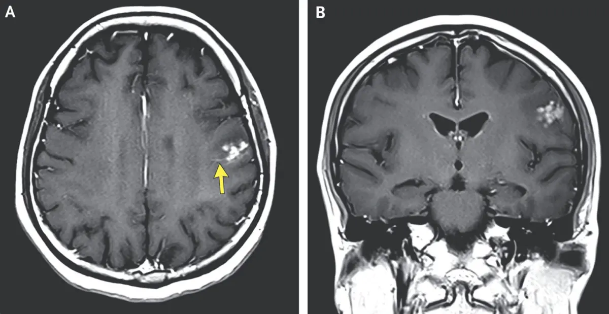
However, given the striking and acute nature of her symptoms, which closely resembled a focal motor seizure, the medical team ordered advanced imaging to evaluate her central nervous system. Magnetic resonance imaging (MRI) of her head provided a swift and definitive explanation for the sudden event.
The brain scans revealed a bright cluster of distinct nodules surrounded by a notable halo of fluid accumulation—known as cerebral edema—located within the left frontal lobe of her brain. Because the left frontal lobe houses critical motor pathways that regulate voluntary movement on the right side of the body, the localized inflammation and tissue pressure caused by these abnormal structures provided a direct anatomical cause for the brief, violent shaking of her right arm and leg.
The characteristic visual appearance of the lesions on the MRI led the medical team to an immediate clinical suspicion regarding their origin: the bright cluster did not appear to be a primary brain tumor or a typical bacterial abscess, but rather a collection of parasite worm eggs embedded deep within her brain tissue.
The detailed account of this unusual medical presentation was recently published in a case report in the New England Journal of Medicine. According to the authors of the report, the patient was diagnosed with suspected neuroschistosomiasis, an uncommon and severe central nervous system complication caused by an infection with a Schistosoma parasitic worm, commonly referred to as a blood fluke.
In the Philippines, where the patient resides and works, the primary endemic blood fluke species responsible for such infections is Schistosoma japonicum. The parasite is also endemic to parts of mainland China and select regions within Indonesia. Despite its scientific name suggesting a connection to Japan, targeted public health interventions, vector control, and environmental management programs have successfully eliminated S. japonicum from the Japanese islands.
An Unfortunate Fluke
The journey of Schistosoma japonicum from an open aquatic environment to the human brain is governed by a complex and diabolical life cycle that relies on specific environmental conditions and multiple host organisms to survive and reproduce.