It began without any of the typical warning signs that usually precede a major neurological event. A woman working as a rice farmer in the Philippines was going about her day feeling entirely healthy—she had experienced no headache, no cognitive confusion, and no generalized body weakness. Then, completely out of nowhere, her right arm and right leg began to shake violently and uncontrollably. For about a minute, half of her body juddered as the unprovoked episode ran its course before the involuntary movements finally subsided.
Recognizing the alarming nature of the sudden motor spasm, the woman sought immediate medical care at a local hospital emergency department. Upon her arrival, attending physicians conducted a standard physical and neurological evaluation. To their initial surprise, the physical examination revealed nothing out of the ordinary; the patient appeared physically intact, exhibiting no persistent focal weakness or obvious signs of systemic illness.
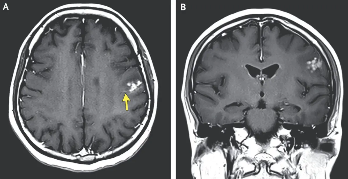
However, given the striking and acute nature of her symptoms, which closely resembled a focal motor seizure, the medical team ordered advanced imaging to evaluate her central nervous system. Magnetic resonance imaging (MRI) of her head provided a swift and definitive explanation for the sudden event.
The brain scans revealed a bright cluster of distinct nodules surrounded by a notable halo of fluid accumulation—known as cerebral edema—located within the left frontal lobe of her brain. Because the left frontal lobe houses critical motor pathways that regulate voluntary movement on the right side of the body, the localized inflammation and tissue pressure caused by these abnormal structures provided a direct anatomical cause for the brief, violent shaking of her right arm and leg.
The characteristic visual appearance of the lesions on the MRI led the medical team to an immediate clinical suspicion regarding their origin: the bright cluster did not appear to be a primary brain tumor or a typical bacterial abscess, but rather a collection of parasite worm eggs embedded deep within her brain tissue.
The detailed account of this unusual medical presentation was recently published in a case report in the New England Journal of Medicine. According to the authors of the report, the patient was diagnosed with suspected neuroschistosomiasis, an uncommon and severe central nervous system complication caused by an infection with a Schistosoma parasitic worm, commonly referred to as a blood fluke.
In the Philippines, where the patient resides and works, the primary endemic blood fluke species responsible for such infections is Schistosoma japonicum. The parasite is also endemic to parts of mainland China and select regions within Indonesia. Despite its scientific name suggesting a connection to Japan, targeted public health interventions, vector control, and environmental management programs have successfully eliminated S. japonicum from the Japanese islands.
An Unfortunate Fluke
The journey of Schistosoma japonicum from an open aquatic environment to the human brain is governed by a complex and diabolical life cycle that relies on specific environmental conditions and multiple host organisms to survive and reproduce.
The transmission cycle begins when parasite eggs are shed into the environment through the urine or feces of infected mammalian hosts, which include humans as well as various domestic and wild animals. In rural agricultural settings, particularly where sanitation infrastructure may be limited, these excreted eggs frequently contaminate nearby freshwater bodies, drainage ditches, and flooded agricultural fields.
When the eggs land in suitable freshwater conditions, they hatch into a free-swimming, microscopic larval form. These aquatic larvae do not immediately infect human hosts; instead, they are biological obligates that must first locate specific species of freshwater snails that serve as intermediate hosts. Upon identifying a suitable snail, the larva actively bores through the mollusk’s outer tissue to gain entry. Inside the soft tissues of the snail, the parasite undergoes two distinct developmental life stages, reproducing asexually and multiplying significantly in overall number.
After completing this essential phase of development within the snail, late-stage larvae burst out of the intermediate host and re-enter the surrounding water column. At this stage, the larvae are known as cercariae. Physical characteristics of cercariae include a distinctive forked tail that aids in their propulsion through the water as they swim in search of a definitive mammalian host.
Unlike many other waterborne parasites that must be ingested to cause infection, cercariae possess the extraordinary biological capability to tunnel directly through intact host skin upon contact. This mode of transmission makes individuals who spend prolonged periods standing or working in freshwater particularly vulnerable to infection.
For a rice farmer in the Philippines, daily agricultural duties often involve wading barefoot or in thin clothing through flooded rice paddies. When cercariae present in the irrigation water encounter exposed human skin, they attach to the surface, shed their forked tails, and utilize specialized chemical secretions to penetrate the outer layers of the skin.
Once the larvae successfully penetrate the skin, they enter the host’s vascular system, initiating a complex migration through the cardiovascular network. The young parasites travel through the venous blood vessels to the right atrium and ventricle of the heart, pass through the capillary beds of the lungs, loop back through the left side of the heart, and are swept into the systemic arterial circulation.
Eventually, they navigate into the portal venous system of the liver. It is within the blood vessels of the liver that the immature flukes feed on host blood components, mature, and differentiate into adult male and female worms.
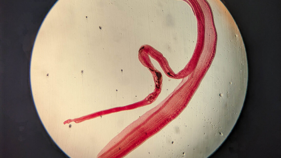
The structural anatomy and behavioral biology of mature Schistosoma worms are notably unique among parasitic flatworms. Adult male and female flukes live permanently paired together in a continuous biological embrace. The thicker adult male parasite features a deep, longitudinal groove along its body known as the gynaecophoric canal. The slender, elongated female worm tucks herself snugly inside this groove, where the coupled pair remains joined throughout their adult lives.
Once mature and paired, these cuddly couples migrate together against the normal flow of blood, exiting the liver to take up residency within the larger blood vessels of the abdomen—specifically the mesenteric veins that connect the liver to the lower gastrointestinal tract. From these abdominal vascular strongholds, the adult pairs feed and begin generating massive quantities of eggs.
Under typical circumstances, the female worm spews out eggs into the tiny venules of the intestinal wall. A substantial portion of these eggs migrate through the mucosal lining of the bowel and are excreted from the host’s body in feces, thereby perpetuating the parasite’s transmission cycle in the environment.
However, because the blood vessels of the mesenteric system are directly connected to systemic circulation pathways, a portion of the produced eggs can be swept backward or carried away by blood flow. In the case of Schistosoma japonicum, the eggs are relatively small and round, making them particularly capable of migrating through small blood vessels and embolizing to distant organ systems.
When parasite eggs escape into the general circulatory network, they can occasionally bypass normal physiological barriers and lodge in ectopic locations, including the central nervous system. Once an egg becomes trapped within the fine blood vessels of the brain tissue, it triggers a potent inflammatory reaction from the host’s immune system.
The immune response attempts to wall off the foreign parasitic material by forming granulomatous nodules around the eggs. In the brain, this localized tissue reaction generates surrounding fluid accumulation, or edema, which exerts structural pressure on adjacent brain tissue.
When this process occurs in the motor regions of the left frontal lobe, as was the case for the Filipino farmer, the localized inflammation and swelling disrupt normal electrical activity in the brain. This acute cortical irritation triggers sudden, involuntary electrical discharges, resulting in the abrupt onset of focal motor seizures that cause the opposite side of the body to shake uncontrollably.
The case presented in the New England Journal of Medicine underscores the complex diagnostic challenges posed by tropical parasitic infections when they manifest in non-typical organs. While schistosomiasis primarily affects the gastrointestinal and hepatic systems in endemic regions, clinicians evaluating patients with sudden, unexplained neurological symptoms must remain vigilant regarding the possibility of central nervous system involvement—particularly when treating individuals with high occupational exposure to freshwater environments.