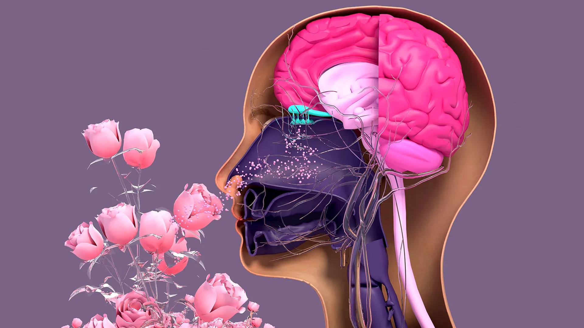
In the complex landscape of the human sensory system, the sense of smell often occupies a paradoxical position. It is frequently taken for granted until it vanishes, a phenomenon that became a global concern during the COVID-19 pandemic. Unlike the delicate neurons of the brain and spinal cord, which possess a notoriously limited capacity for repair, the sensory neurons located within the nasal cavity are among the most resilient and regenerative tissues in the mammalian body. Throughout a person’s life, these neurons are constantly dying and being replaced, a necessary adaptation for cells that are perpetually exposed to the harsh outside environment, including pollutants, allergens, and pathogens.
However, this regenerative engine is not invincible. Factors ranging from viral infections and toxic exposure to the inevitable process of aging can slow or halt the production of new neurons, leading to anosmia—the total loss of smell—or hyposmia, its partial loss. To better understand how this regenerative process functions and why it sometimes fails, researchers at Tufts University School of Medicine and the Graduate School of Biomedical Sciences (GSBS) have developed a sophisticated new three-dimensional model of olfactory tissue.
Their findings, published recently in the journal Cell Reports Methods, have already begun to shift the scientific understanding of nasal biology. Most notably, the study reveals that a specific type of stem cell, long dismissed by many in the field as largely dormant, actually plays a pivotal role in maintaining the health and renewal of our smell-sensing apparatus.
A New Perspective on "Dormant" Stem Cells
For years, the scientific consensus regarding the olfactory epithelium—the specialized tissue inside the nose responsible for detecting odors—centered on two primary types of stem cells: horizontal basal cells (HBCs) and globose basal cells (GBCs). Traditionally, GBCs were viewed as the primary workhorses of the system, the active "progenitor" cells that frequently divide to replace aging or damaged neurons. In contrast, HBCs were often categorized as a reserve population—dormant cells that remained quiet unless a catastrophic injury occurred.
The Tufts research team, led by senior author Brian Lin, a research assistant professor in the Department of Developmental, Molecular and Chemical Biology, has now challenged this "dormant" label. By utilizing their newly devised 3D organoid model, the team discovered that HBCs are far more active and essential than previously realized.
"Our research suggests that these two stem cells may be interdependent," explains Lin. "One type that we thought was largely dormant—HBCs—may actually play a crucial role in supporting the production of new neurons and the repair of damaged tissue."
Through the use of the 3D mouse-tissue model, the researchers identified a specific subpopulation of HBCs characterized by the production of a protein known as KRT5. They observed that these KRT5-producing cells do not just sit idly by; instead, they appear to be vital architects of the regenerative process. When the team experimentally depleted these specific HBCs from the organoid cultures, the results were stark: the generation of new sensory neurons was significantly impaired. This suggests that the HBCs provide a necessary support structure or biochemical signaling environment that allows the GBCs to function effectively. Without the "dormant" cells, the "active" cells cannot complete their mission.
The Power of the 3D Organoid Model
The breakthrough was made possible by the development of an "organoid"—a simplified, miniature version of an organ grown in a laboratory setting that mimics the three-dimensional architecture of actual biological tissue. While traditional two-dimensional cell cultures (cells grown in a flat petri dish) have been the backbone of biological research for decades, they often fail to capture the complex interactions that occur in a living body.
In the nose, cells do not exist in isolation; they are stacked and interwoven, communicating through physical contact and chemical signals. The Tufts team’s 3D model allows researchers to observe these interactions in real-time. This model is particularly effective at showing how HBCs and GBCs communicate and support one another to develop new smell-sensing nerve tissue.
"We wanted to develop an easy-to-use model so that non-stem cell biologists and those working in labs with limited resources could use it to better understand how olfactory neurons regenerate," says Lin.
The accessibility of the model was a primary goal for the study’s lead author, Juliana Gutschow Gameiro. A former Ph.D. student visiting GSBS from the State University of Londrina in Brazil, Gameiro was dedicated to ensuring the model could be replicated in laboratories that might lack the massive funding or high-end equipment of elite research institutions. This "democratization" of the research tool is particularly timely, as interest in the olfactory system has surged in the wake of the pandemic.
"Because loss of smell is associated with COVID-19, as well as with Parkinson’s disease and other conditions, a much larger number of researchers from a variety of different fields have begun researching olfactory epithelial cells in the last few years," Lin notes. By providing a low-cost, high-fidelity model, the Tufts team is opening the door for scientists worldwide to investigate why the sense of smell diminishes or fails completely in various patient populations.
Investigating the Impact of Aging
One of the most pressing questions in olfactory research is why our sense of smell tends to decline as we age. It is a common clinical observation that older adults often lose the ability to detect subtle scents, which can impact everything from their appetite and nutrition to their ability to detect environmental hazards like gas leaks or spoiled food.
Using their new model, the Tufts researchers compared cells harvested from young mice with those from older mice. They found a definitive decline in the ability of the older cells to generate new neurons within the organoid environment.
"We found a decline in the ability of the older mice cells to generate new neurons," Lin says. "We think this is due to a decrease in the GBC population as we age, but we need to do more work to test this hypothesis and if so, develop ways to rejuvenate them."
The discovery that the GBC population might be dwindling with age provides a specific target for future therapies. If scientists can find a way to "reboot" these stem cells or supplement the aging tissue with the supportive signals provided by the HBCs, it might be possible to restore a youthful level of regenerative capacity to the aging nose.
The Path Toward Human Treatment
While the current research relies on a mouse model, the ultimate objective of the Tufts team is far more ambitious: the creation of a human olfactory organoid. Such a model would be a transformative tool for modern medicine, serving as a platform for screening potential drugs to treat patients suffering from chronic smell loss.
Organoids offer a distinct advantage in drug development. They are quicker and less expensive to work with than whole-animal models, and they can be more effective than traditional human cell cultures because they more accurately represent human physiology. While organoids have been successfully developed for other organs, such as the lungs and kidneys, the human olfactory system has proven to be a uniquely difficult challenge.
"It’s challenging to get pure olfactory tissue from humans," explains Lin. The process of collecting these cells is invasive and technically difficult. Currently, a procedure similar to a COVID-19 swab is used, where a brush is pushed deep into the nasal cavity of an anesthetized patient. However, the resulting sample is often a mixture of different cell types.
Unlike the mouse model, where researchers can easily isolate the desired stem cells, human samples collected via nasal brushing often contain a mix of respiratory stem cells and olfactory stem cells. Distinguishing and separating these two types is a significant hurdle. If respiratory cells dominate the culture, the organoid will not function as a true model of the smell-sensing tissue.
The research team’s next major challenge is to refine a simple, inexpensive technique to separate these human olfactory stem cells and "coax" them to grow in the laboratory. Once a stable human organoid is achieved, it could be used to test a wide array of pharmaceutical compounds, looking for those that can trigger the HBCs and GBCs to begin the work of repair.
A Growing Field of Study
The implications of this research extend beyond the immediate recovery of smell for post-viral patients. The olfactory system is increasingly viewed by scientists as a window into the brain. Because the olfactory neurons are directly connected to the brain’s olfactory bulb, the nose is one of the few places where the nervous system is in direct contact with the external world.
There is a well-documented link between a declining sense of smell and the early stages of neurodegenerative diseases like Alzheimer’s and Parkinson’s. By understanding the fundamental mechanics of how nasal nerve tissue regenerates—and why that regeneration fails—researchers may gain broader insights into the regenerative limitations of the central nervous system as a whole.
For now, the Tufts study provides a vital piece of the puzzle. By identifying the critical role of the "dormant" horizontal basal cells and providing a new, accessible tool for study, the team has laid the groundwork for a new era of olfactory research. As the scientific community continues to grapple with the long-term sensory impacts of the modern environment and aging, the humble stem cells of the nose may hold the key to restoring one of our most essential connections to the world around us.