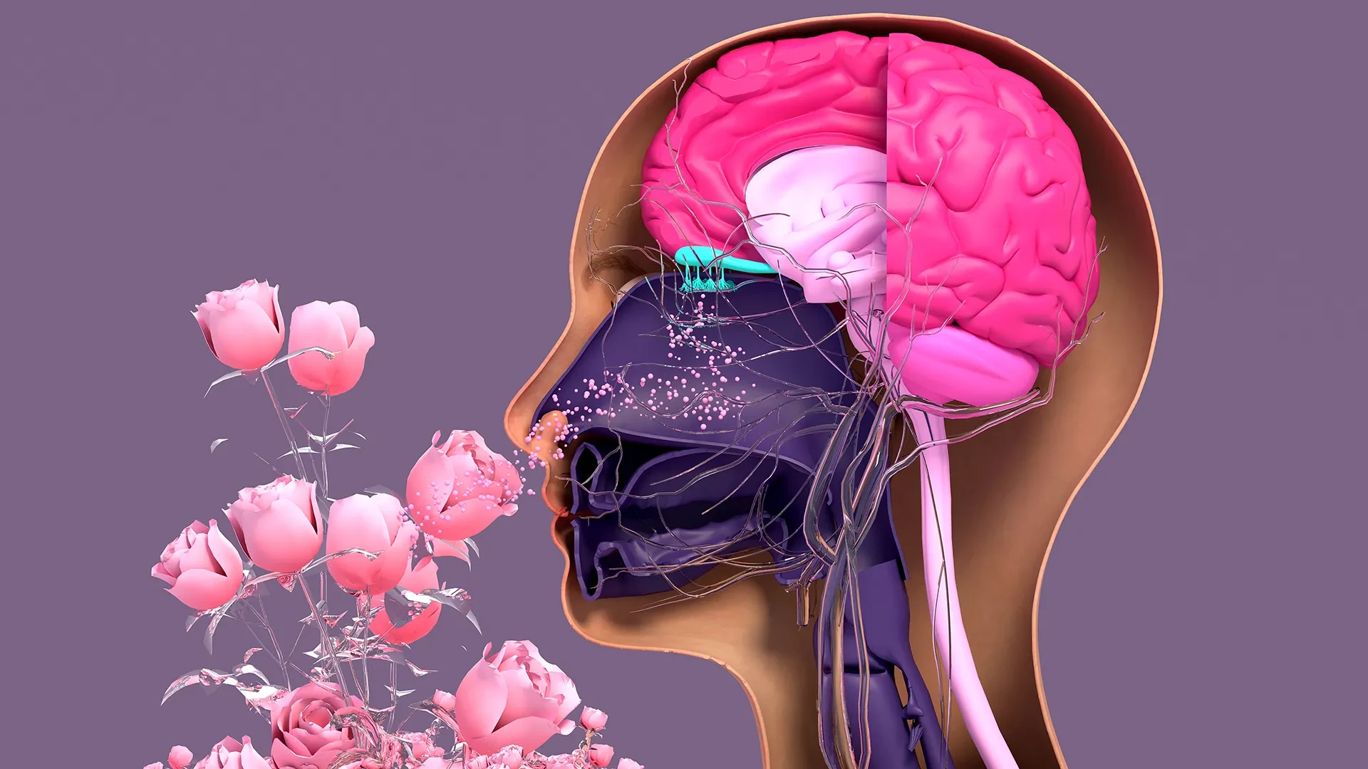
In the intricate landscape of human biology, the sense of smell often occupies a paradoxical position. While it is frequently regarded as the most visceral and evocative of our five senses—capable of triggering distant memories or alerting us to immediate danger—it is also one of the most vulnerable. Unlike the protected confines of the central nervous system, the sensory neurons responsible for detecting odors are situated in the nasal cavity, where they are relentlessly exposed to the external world. Every breath draws in a cocktail of dust, pollutants, allergens, and pathogens, placing these delicate nerve tissues under constant siege.
Despite this environmental bombardment, the olfactory system possesses a remarkable, almost unique, capacity for self-renewal. Throughout a person’s life, these neurons are replaced and repaired in a cycle of regeneration that usually keeps the sense of smell sharp. However, this process is not invincible. Factors such as viral infections—most notably COVID-19—exposure to environmental toxins, and the natural progression of aging can severely hamper this regenerative ability, leading to anosmia or hyposmia, the total or partial loss of smell.
Now, a pioneering study from researchers at Tufts University School of Medicine and the Graduate School of Biomedical Sciences (GSBS), in collaboration with international colleagues, has shed new light on how this regeneration occurs. By developing a sophisticated three-dimensional "organoid" model of nasal tissue, the team has discovered that a specific type of stem cell, long dismissed as largely dormant, actually plays a pivotal role in maintaining and repairing our olfactory machinery. Their findings, recently published in the journal Cell Reports Methods, offer a new roadmap for understanding sensory loss and potentially developing future treatments to restore it.
The Mystery of the Nasal Stem Cell
At the heart of the study are two primary types of stem cells found within the olfactory epithelium, the specialized tissue lining the nasal cavity: horizontal basal cells (HBCs) and globose basal cells (GBCs). For years, the scientific consensus held a somewhat binary view of these cells. The GBCs were seen as the "workhorses" of the system—actively dividing and differentiating into new sensory neurons to replace those that had died. In contrast, the HBCs were viewed as a reserve population, a "break glass in case of emergency" backup that remained largely inactive under normal conditions.
However, the Tufts research team, led by senior author Brian Lin, a research assistant professor in the Department of Developmental, Molecular and Chemical Biology, found that this traditional view was incomplete. Using their newly devised 3D mouse model, the researchers observed a complex, interdependent relationship between the two cell types that had previously been invisible in simpler 2D cultures.
"Our research suggests that these two stem cells may be interdependent," says Lin. "One type that we thought was largely dormant—HBCs—may actually play a crucial role in supporting the production of new neurons and the repair of damaged tissue."
By observing the cells in a 3D environment that more closely mimics the natural architecture of the nose, the team identified a specific subpopulation of HBCs. These cells are distinguished by their production of a protein known as KRT5. Rather than sitting idly by, these KRT5-positive HBCs were found to be active participants in the regenerative process. They appear to act as a support system, creating the necessary environment and biochemical cues for the GBCs to successfully generate new olfactory neurons.
To test the importance of these cells, the researchers performed a depletion experiment. When the KRT5-producing HBCs were selectively removed from the organoid cultures, the results were striking: the generation of new neurons was significantly impaired. This suggested that without the support of these "dormant" cells, the more active GBCs could not fulfill their role effectively. The discovery elevates the HBC from a passive bystander to an essential player in the maintenance of human olfaction.
Aging and the Decline of Sensory Renewal
Beyond identifying the key players in regeneration, the study also delved into why our sense of smell tends to fade as we get older. It is a common clinical observation that elderly individuals often experience a diminished ability to detect odors, which can impact nutrition, safety, and overall quality of life.
The research team utilized their 3D model to compare the regenerative capacities of cells harvested from mice of different ages. They found a clear correlation between age and a decline in the ability to produce new neurons.
"We found a decline in the ability of the older mice cells to generate new neurons," Lin notes. "We think this is due to a decrease in the GBC population as we age, but we need to do more work to test this hypothesis and if so, develop ways to rejuvenate them."
The implications of this finding are significant. If the loss of smell in aging is driven by a depletion of the GBC population or a breakdown in the communication between HBCs and GBCs, it may be possible to develop therapeutic interventions. The goal would be to "reboot" these stem cell populations, essentially tricking the nasal tissue into a more youthful state of high-turnover regeneration.
An Easy-To-Use Model
One of the most significant contributions of this research is not just the biological discovery itself, but the tool used to achieve it. The 3D olfactory organoid developed by the team was designed with a specific philosophy in mind: accessibility.
The lead author of the study, Juliana Gutschow Gameiro, was a visiting Ph.D. student at GSBS from the State University of Londrina in Brazil. Her presence at Tufts was driven by a desire to create a research model that did not require the astronomical budgets or high-end specialized equipment often found in elite Western institutions.
"Because loss of smell is associated with COVID-19, as well as with Parkinson’s disease and other conditions, a much larger number of researchers from a variety of different fields have begun researching olfactory epithelial cells in the last few years," says Lin.
The sudden global focus on anosmia during the pandemic highlighted a gap in the available research tools. While many labs wanted to study the nasal epithelium, many were deterred by the complexity and cost of maintaining primary cell cultures or animal models. By creating an "easy-to-create" 3D organoid, Gameiro and the Tufts team have democratized this area of study.
"We wanted to develop an easy-to-use model so that non-stem cell biologists and those working in labs with limited resources could use it to better understand how olfactory neurons regenerate and what happens that causes that process to diminish or fail completely," Lin explains.
This model allows researchers to grow "mini-noses" in a dish, providing a controlled environment to test how various toxins, viruses, or drugs affect the stem cells and their ability to produce neurons. Because it is 3D, it captures the physical interactions between different cell layers, a critical factor that 2D Petri dish models often fail to replicate.
Next Step: A Human Organoid
While the mouse model has provided a wealth of new information, the researchers are already looking toward the ultimate prize: a human olfactory organoid. The transition from animal models to human tissue is a standard but difficult hurdle in biomedical research.
The potential for a human version of this model is vast. It would allow scientists to perform high-throughput screening of drugs to treat people suffering from chronic smell loss. Currently, treatments for anosmia are limited and often involve corticosteroids, which may not address the underlying failure of the stem cell population. A human organoid could be used to find compounds that specifically stimulate HBCs or GBCs to resume their regenerative work.
Furthermore, organoids represent a more ethical and efficient alternative to traditional research. They can make pre-clinical trials quicker and less expensive than using whole animal models, and they are often more accurate than existing human cell lines that have been modified to grow indefinitely in a lab.
However, creating a human olfactory organoid presents unique logistical challenges. "It’s challenging to get pure olfactory tissue from humans," Lin says.
In a clinical setting, obtaining these cells requires a procedure where an individual is anesthetized, and a brush—similar to the swabs used for COVID-19 testing but designed to reach much deeper—is inserted into the uppermost part of the nasal cavity. The primary difficulty lies in the fact that the human nose contains a mix of respiratory tissue (which helps us breathe) and olfactory tissue (which helps us smell). In the samples collected via brushing, the stem cells from these two different types of tissue are often mixed together and are notoriously difficult to separate.
The Tufts research team’s next major challenge is to refine their techniques to isolate these human olfactory stem cells from their respiratory counterparts. Once separated, they must then find the right "recipe" of nutrients and growth factors to coax them into forming a 3D organoid in the lab.
The successful development of a human model would bridge the gap between basic laboratory science and clinical treatment. For millions of people worldwide whose lives have been dampened by the loss of smell—whether through the long-term effects of a virus, the progression of neurodegenerative disease, or simply the passage of time—this research offers a glimmer of hope that the "dormant" healers within their own bodies might one day be awakened.