The Nikon Small World in Motion competition, a prestigious annual event that celebrates the intersection of high-level scientific research and breathtaking visual artistry, has been thrust into a state of controversy. The scientific community is currently reeling from allegations that the most recent first-place winner, Dr. Ning Xu, may have utilized prohibited artificial intelligence tools to create his award-winning entry. What began as a celebration of microscopic discovery has rapidly transformed into a rigorous investigation into the integrity of digital imaging in the age of generative AI, prompting a formal review by Nikon and sparking a heated debate among researchers worldwide.
For decades, the Nikon Small World competition has served as the premier showcase for the "hidden world" that exists beyond the reach of the naked eye. While the photography category has been a staple since 1975, the Small World in Motion category, introduced more recently, focuses on the dynamic, living processes of biology captured through time-lapse and high-speed microscopy. The entries are typically abstract, often appearing as alien landscapes or mesmerizing patterns to the layperson, but they are rooted in the rigorous demands of biological science. However, when Dr. Xu, an optical engineering researcher at the National University of Singapore, was announced as the winner last month, the initial awe was quickly replaced by skepticism from experts who noticed anomalies that seemed to defy the laws of biological physics.
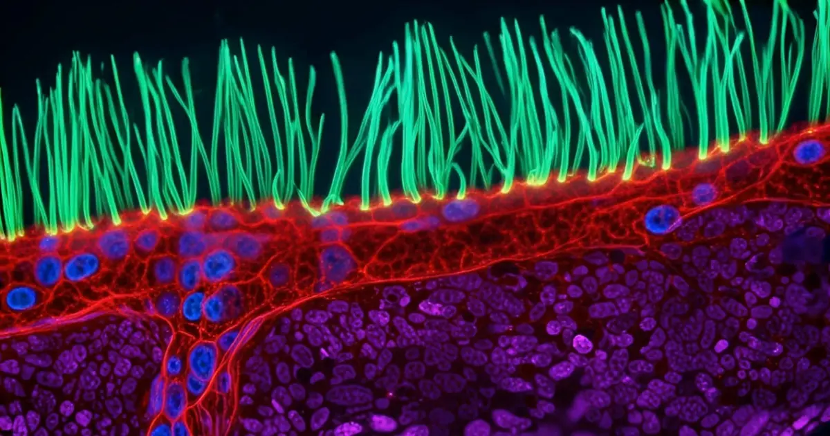
The winning video was intended to provide a rare glimpse into the movement of cilia—tiny, hair-like structures that line the respiratory tract and play a critical role in clearing mucus and foreign particles from the lungs. According to the submission details provided by Dr. Xu, the tissue used for the imaging was obtained from a child suffering from primary ciliary dyskinesia (PCD). This rare genetic disorder impairs the function of the cilia, leading to chronic respiratory issues, including persistent lung, sinus, and ear infections. Capturing the motion of these structures in such a specific pathological context was hailed as a significant achievement, both for its visual complexity and its potential scientific value.
The acclaim was short-lived. Almost immediately following the announcement, members of the scientific community began to voice their concerns publicly, pointing toward what they described as "continuity errors" that are uncharacteristic of authentic microscopy. Edward Phelps, an associate professor at the University of Florida, was among the first to raise a red flag. Writing on LinkedIn, Phelps detailed several "serious problems" within the footage that suggested the video was not a pure representation of captured data.
Phelps observed that certain "purple structures" within the frame appeared to "pop in and out of existence," a phenomenon that does not occur in standard fluorescence microscopy unless there is a catastrophic failure of the sample or the equipment. Furthermore, he noted that the green cilia appeared to materialize from nowhere, exhibiting movements and sizes that did not align with established biological measurements. In the precise world of microscopy, where structures are measured in micrometers, such discrepancies are often the first sign of digital manipulation or synthetic generation.